So, your practice recently purchased or is now considering a dental microscope. As a surgical assistant, this could be a revolution in how you and your surgeon diagnose, create treatment plans, conduct procedures, review post-op and even more. Plus, it could improve your communication during operations, as you both now have the ability to see into the oral cavity at the same time, at higher magnification levels.
The procedures you offer may require multiple people on your team to be able to view your patient’s anatomy. But, this can be challenging when viewing your patient’s oral cavity, due to its relatively small size and dark shadows. Unlike an X-ray or CBCT scan, which captures a single moment, seeing the anatomy in real-time, at the same time, can be critically important for surgeons, assistants and staff.
This is something uniquely powerful about the dental microscope over any other magnification technology. There are numerous features found on modern dental microscopes that surgical assistants, hygienists and other staff members should know about in order to maximize the value of their scope, and increase the efficacy of their exams and practice overall.
Today, we’re going to cover these features, as well as the leading benefits of a scope for surgical assistants and staff members.
Video Monitors

One of the most common ways doctors, staff and patients can collectively view the oral cavity is through installing a video camera and monitor. The video monitor can become an essential communication tool between the doctor using the scope and all staff and assistants.
With a live-feed, everyone present in the operatory can view the doctor’s actions firsthand, and anticipate their movements. Some doctors we speak with say this dramatically improves their communication with staff, because it reduces their need for verbalizing each action, thereby making their surgeries more efficient.
In order to maintain optimal viewing, make sure your monitor or monitors are strategically positioned in your operatory. When placing a monitor in your operatory, we advise against placing just where there is open space on a wall. Over the course of the work day, your body may become fatigued from craning your head and neck to view the screen. Because one of the leading benefits of a scope is improved ergonomics, it’s important everyone on staff gets this benefit – thereby extending the health of your practice.
Instead, we recommend strategically placing monitors straight across from whoever will be looking at the monitor during exams. Some doctors will use multiple monitors in their operatories: one used by you, the surgical assistant, one directly in front of the doctor to ensure the camera is centered for proper documentation, and one on the ceiling for the patient to view if they desire. In this configuration, your practice is maximizing the visualization of the scope, reaping the benefits of improved ergonomics, procedural communication, staff training, clinical efficiency, and patient education – all at once!
We also found that using a Dual Iris Diaphragm in conjunction with your camera helps achieve a greater depth of field for their live-feeds and recordings.
What does a Dual Iris Diaphragm do?
Our eyes are designed to see in stereoscopic vision, meaning our brains are processing two different visuals from each eye, then stitching the visuals together in a three-dimensional view. A camera flattens an image to what would be seen by a single “eye”, meaning you aren’t seeing the same depth perception as you would in an analog view from the scope (or even with the naked eye).
The Dual Iris Diaphragm solves for this by providing a more true-to-life visual, increasing the depth of field to both the user and any camera attached. It is mostly used with DSLR style cameras.
For more information on how our eyes and vision work while using magnification, check out our article: How is Magnification Used in Dentistry? Why the Naked Eye Isn’t Always Enough.
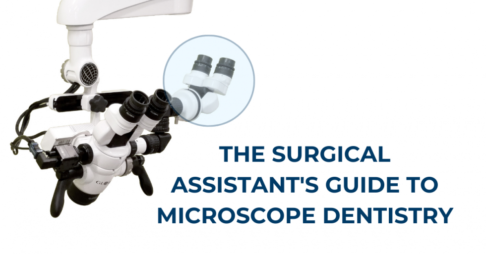
Binocular Co-Observation System

Another popular feature we recommend to doctors working with surgical assistants is a Binocular Co-Observation System (BOS). As the name suggests, this attachment adds a second pair of binoculars off of the primary scope. This allows a second operator to be directly involved in a case, seeing the fine detail the primary user sees. For some operatories, this second binocular vision is preferable to viewing through a screen or monitor because it allows closer involvement from an assistant. You’ll be in immediate proximity to the patient’s oral cavity, giving you the ability to work without limiting your vision or looking at a monitor.
A BOS can be configured in either left or right position, depending on which is best suited for your unique operatory setup. The BOS pulls light from either the left or right side of the scope depending on which side it is installed on. This limits the field of view on the second binocular to pseudo-stereo vision, which is not as wide as what the primary user will see with both eyes to form the image. Otherwise, a BOS provides the magnification, illumination and analog viewing to help surgical assistants work alongside the primary user to conduct treatments.
Mounting Options
One final consideration when configuring your operatory with a microscope is your mounting selection. As a surgical assistant, it’s important to get familiar with how and where you and your staff will be positioned in the operatory while using the scope.
At Global Surgical, we offer more mounting options than any other brand to help you get the best possible configuration. These options include:
- Ceiling mounts for ceilings 8-10 ft
- High and low wall mounts
- Floor stands
- Mobile floor stand (a rolling cart configuration for transportation between operatories)
For more on this topic, and a full list of mounting options (including diagrams) available through Global Surgical, continue reading: How to Mount a Dental Microscope.
BONUS: Micro-Assistant’s Chair

One addition to keep in mind outside of magnification and and optimal viewing is your comfort and long-term ergonomics. While the dental microscope was designed over 25 years to help improve ergonomics, there are additional tools like the Micro-Surgeon’s chair which can further alleviate back and neck strain.
A fully adjustable Micro-Surgeon’s Chair offers proper support for your lumbar area, as well as forearms, so you can perform fine motor skills in a completely relaxed position. Ergonomic chairs like these come in varying designs and shapes, so be sure to find a chair which is adjustable to your height and size.
Here at Global Surgical, in addition to our A-Series microscopes, we also manufacture Micro-Surgeon’s and Micro-Assistant’s chairs in our US facility to give you a complete ergonomic solution for dental surgeries. Our Micro-Assistant’s chair differs slightly from our Micro-Surgeon’s chair with a special curved armrest for flexible positioning, comfort and stability for the assistant. Its adjustability helps to prevent repetitive stress injuries by providing all of the features of the Micro-Surgeon’s Chair.
Interested in adding a Micro-Assistant’s chair to your practice? Give us a call 800-861-3585 or by completing an online form here.
Benefits of a Dental Microscope for a Surgical Assistant
Now that we’ve covered many of the common features doctors choose to support their assistants and staff, let’s review the top 3 benefits you can expect from your new scope.
#1 Communication
The oral cavity is unique from other parts of the body because of its relatively small size and dark shadows. As such, it can be challenging to share a view with more than one person at a time. As we’ve discussed, tools like a video monitor and a BOS can dramatically improve communication because multiple people can view at once. Imagine being able to see what the doctor sees, with a high power of magnification, without needing to flip between devices to get a real-time visual.
With this visualization, you can process non-verbal queues from the doctor by anticipating movement, making your work more efficient. With a video monitor set up, you can also use these visuals to improve staff training as well as use it as a tool for patient communication. Like an intraoral scan or CBCT image, you can display your patient’s anatomy for true chairside visualization. This improves your ability to educate the patient, and leads to greater treatment acceptance.
#2 Documentation
Next, as a surgical assistant, part of your role may involve case documentation. Outside of case work, adding video functionality to your microscope (typically with a DSLR camera or similar HD video source) is also useful for submitting insurance reimbursements and adding video to clinical cases/lectures and patient education. Just like an X-ray or intraoral scan, these video files or photo stills can be exported and attached to your patient’s records.
#3 Training & Education
When we were talking about communication, we mentioned how effective a dental microscope can be for training and education purposes. The reason these are so critical to you is because training and education impact the “bottom line” for a practice.
Training – Any staff present in the exam room with a microscope video monitor will have the ability to see dental surgeries first-hand. As staff members hear discussions between you and your doctor, they will be more familiar with the ways you communicate and the resources you’ll require. With more active staff training around a microscope, your staff will also be prepared to care for the scope, such as performing routine cleaning and preventative maintenance. This training, in turn, can help your practice maximize the lifespan of your scope and improve efficiencies between exams.
For more information on these topics, continue reading our help guides:
Education – With greater documentation captured by a camera attachment, you’ll be able to build a stronger, more compelling case file to use for educational purposes. Patient education leads to greater treatment acceptance, helping you work more cases and increase your revenue.
Visualization at a high level of magnification helps ensure you’re capturing all the details when providing a diagnosis. Precision is key to diagnosis as well as planning treatment options. And, when effectively communicated to your patients, maximizes the benefit of using a dental microscope.
To explore more of the top ways dental professionals use magnification to benefit their practice, continue reading: Top 5 Uses for a Dental Microscope.
More Value With a Global Microscope
At Global Surgical, we’ve been committed to building microscopes and providing exceptional service for over 25 years. We’ve worked with thousands of dental professionals to help them get the most value out of their scope. With our experience, we’ve heard from many doctors on how they’ve configured their practice to best suit their staff and patients so they can conduct the best patient care possible.
As part of keeping our commitment to you, the user, we’ve developed several features that help you get the best experience, including offering more mounting options than any other brand. We’re also the only dental microscope manufacturer offering a limited lifetime warranty (US & Canada only), meaning we’ll stand by the quality of the scope you’re using.
As you get more comfortable training with a scope, you’ll be able to reap the benefits of more efficient surgeries with greater confidence in communication, documentation, training and education. Many of the doctors we speak with quickly begin using their scopes with nearly every patient – from observation and diagnosis to treatment, so we know it’s important they are able to be operational every single day for years to come.
But don’t just take our word for it, hear from doctors who have used Global microscopes in their practices:
- “All of my hygienists and my associates use the microscope for all procedures as well. My hygienists are more efficient with the microscope than they ever were with loupes.” – Dr. Wayne Remington, DDS
- “Microscopy is an integral part, not only of how we diagnose and treat our patients, but key to how Team Atlanta teaches – enlarged images and close-up video for effective communication.” – Team Atlanta: Dr. Ronald Goldstein, Dr. Maurice Salama, Dr, David Garber & Dr. Henry Salama
- “[My microscope] has been a wonderful addition to my practice and my patients view it as another example of my dedication to their dentistry.” – Dr. Michael C. McVicker, DDS
Ready to Get Started? Reach Out!
If you’re just getting started with a scope, or considering adding a scope, we are here to help! We can help configure and customize your scope to you, your surgeon and your staff’s needs.
We’re proud to be based in the US, with manufacturing and assembly facilities in St. Louis, MO. This helps us give our customers the best service, domestically and internationally.
Get started by reaching out to us at 800-861-3585 or by clicking the button below.