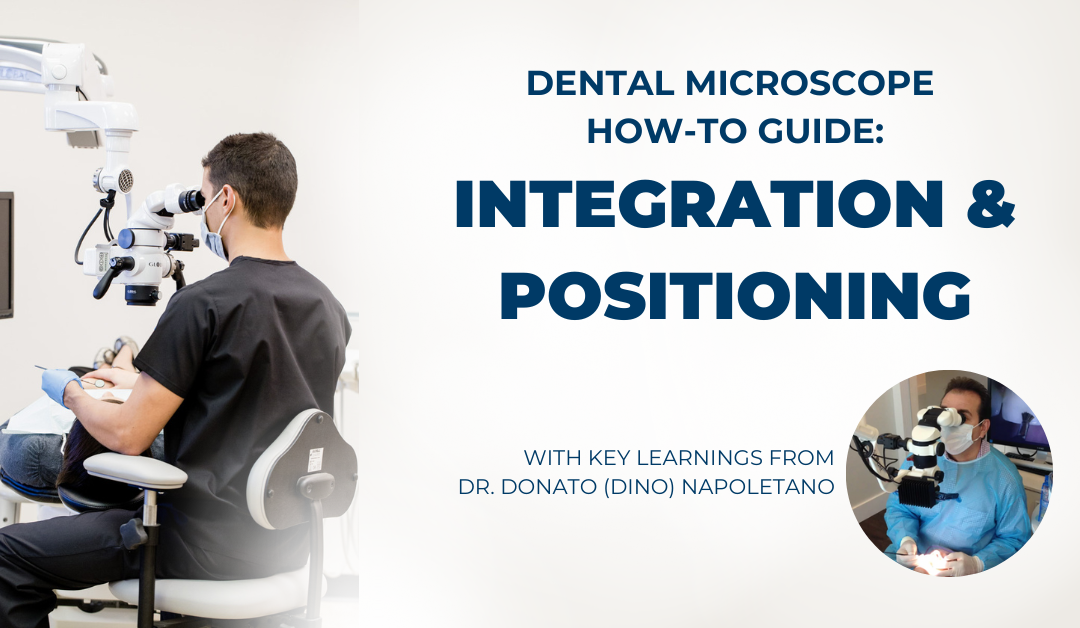
Dental Microscope How-to Guide: Integration & Positioning

For over 25 years, the AAE (American Association of Endodontists) has been strongly advocating for the use of a dental microscope, otherwise known as DOM (dental operating microscope) or surgical microscope, in dental practices as well as during doctoral study.
Today, doctors both specialized in endodontics as well as in general or specialty practice are choosing scopes because of their:
- multiple levels of magnification
- shadow-free illumination
- increased diagnostic capability & precision
- ergonomics & long-term health
- patient education & digital documentation capabilities
Since the dawn of this era of microscopic dentistry, the founders at Global Surgical became the first company to focus on manufacturing dental microscopes. With our years of experience, we’ve worked closely with doctors to help them maximize the value they see from a dental microscope. And, as part of our commitment to our customers, we’ve collected considerable feedback to help us maintain a high standard of excellence. You’ll see this today with both our product and accessory designs, as well as the training, manuals and articles we produce to help new customers get up and running on their scope as efficiently as possible.
As part of this effort, we launched a collection of the most requested reference materials from doctors we work with – our “Dental Microscope How-to Guides”. Each article in this series will include easy-to-follow steps to help reduce the number of training hours on specific areas most commonly referenced by doctors we work with.
In Part 1 in this series, we covered how to focus your dental microscope. In this article, we explained the steps for parfocal adjustment in coarse and fine focus, as well as the use of a multifocal lens attachment. Understanding how to focus your scope is an essential skill for microscopy because every time you switch magnification levels or reposition your patient, you’ll need to adjust your parfocal lens to see clearly.
PRO TIP: Whether or not you’ve trained on a scope, we suggest to typically expect a 4-6 month period of using your scope before you’ll feel comfortable during daily use. As you get more skilled with your scope, you’ll be able to increase the speed and efficiency at which you’ll operate.
Today, we’re offering our expertise on the topic of positioning and integration. Mastering the use of a dental microscope requires getting comfortable with indirect vision. This means, while your vision is enhanced through the binoculars of the scope, you also need to acclimate your hands and body for using your dental instruments as you would with direct vision.
In order to realize the ergonomic and visualization benefits enjoyed by microscope users, getting positioning and integration right is a critical first step.
The following positioning guide is compiled in-part by Dr. Donato (Dino) Napoletano, a restorative dentist and long-time Global microscope user. In addition to owning and operating Donato Dental in Middletown, New York, Dr. Napoletano has conducted hands-on microscope training sessions both privately and at Tufts University’s GPR program.
To get started, let’s review the 3 following basics of integrating your scope into your practice:
Location
This may seem obvious after you read this, but it remains true with many doctors using scopes… if your scope is not positioned or mounted with easy access around your operatory, it will simply not be used. Most dental microscopes can either be permanently mounted to the floor, wall or ceiling or attached on a movable floor stand.
Consult your manufacturer or owner’s manual when planning your scope installation. While every operatory is unique, they’ve likely worked with numerous different configurations to help you maximize your setup. Getting the best configuration for your scope all depends on how much space you have to work with in the room, and what other features in the room need to be configured to. Also, consider how you’ll incorporate audio/visual with a live feed monitor for assistant/staff/patient viewing.
Per the Global Surgical owner’s manual: The most common mounting location for ceiling mount and floor mount models is over the patient’s left hip. A wall mount is most typically mounted on right side.

For more guidance on planning for your dental microscope installation, check out our article: How to Mount a Dental Microscope.
Parfocaling
Parfocaling a scope means tuning the scope to the individual user’s eyesight. This is a simple action covered in the owner’s manual. This should only take a few minutes to do and should be done periodically.
The importance of parfocaling your scope is two-fold.
- It helps to ensure that the microscope stays in relative focus when changing levels of magnification without having to move your scope up or down to bring the operating field back into focus.
- It helps to ensure that images and videos recorded on an integrated digital still camera are in-focus. When your scope is not parfocaled correctly to the operator’s eyes, an image that is in-focus to you, the operator, may be out of focus to the camera.
Also, when you parfocal, ensure you’ve adjusted the diopter to your eyes. This is similar to setting your optics up like an eyeglass prescription, therefore every user has their own settings.
Position
Learning how to effectively position your scope so that the operating field or target tooth can be visualized is part of getting comfortable with your scope. To get acclimated, some doctors seek formal training, while some will learn it through trial and error.
The most important things to be mindful of when learning how to get comfortable with microscope positioning is that in addition to your scope itself, the position of the exam chair, patient’s head and mirror also need to be considered when trying to visualize the operating field or a target tooth.
While mastering positioning will require training and practice in the real-world, we’ve outlined six tips and tricks from doctors to help you bridge the learning gap as efficiently as possible.
#1 Keep the patient chair-height low. Actually, in most cases, there is no need to raise it at all! If you’re accustomed to working with loupes or completely unassisted vision, most doctors tend to recline the chair back and also raise the chair height in order to bring the operating field closer to your eyes. When working with your scope, it is preferable to recline the chair back and not raise the chair height much, if at all.
The reason for this is that if the chair is raised too high, your scope will also need to be positioned higher in order to focus on the operating field. While most dental microscopes are equipped with inclinable binoculars, if your patient chair is too high, even at its most downward position, you won’t be able to comfortably reach the eyepieces. Thus, the poor position of the scope will cause strain and negate some of the ergonomic benefits of sitting comfortably in a rested position. Proper positioning allows you to conduct exams and perform treatments for prolonged periods while still maintaining your long-term health.
For more insight into unassisted vision vs visual enhancement through a scope (including an intriguing review of our eye’s anatomy and limitations), continue reading: How is Magnification Used in Dentistry? Why the Naked Eye Isn’t Always Enough.
#2 Fully recline your patient’s chair and, once reclined, have the patient scoot up on the chair. When you begin reclining your chair back, your patient’s body tends to slouch downward on the chair. This positions their head further away from the operator. So once the chair is reclined, your patient should always be asked to scoot their bodies up on the chair.
This brings your patient’s head closer to you, which in turn allows you to position your scope closer so you won’t need to tilt your head excessively forward to reach the oculars.
TIP FROM DR. NAPOLETANO: Most dental microscope manufacturers have an optional attachment called an extender (which I highly recommend), that extends the eyepieces even closer to the operator, further reducing the amount of forward neck-tilt needed to reach the oculars.
#3 Move your patient’s head around often. One of the biggest ergonomic pitfalls we hear from doctors just starting on a microscope is overcompensating your positioning in an attempt to visualize a target tooth. Many will tend to move, rotate or tilt their scope around in positions that are not ergonomically friendly to the operator.
Remember that in addition to your scope, your patient’s head can move too! Effectively visualizing all areas of the mouth often requires a combination of tilting and rotating your scope, the patient’s head and your mirror.
For example, when trying to visualize maxillary posterior teeth, having the patient tilt their head back (chin-up) will help increase your visibility.
A slight rotation of both the patient’s head and your scope may also be required at times. For instance, when trying to visualize a posterior tooth in the left arch, a slight rotation of your scope and the patient’s head to the left will be helpful (assuming the operator is right-handed and working between 12 o’clock and 9 o’clock position). In our experience, this is the most ergonomically-friendly position to work from when using your scope.
#4 Sit ergonomically upright and position the scope to you. The best way for you to maximize the ergonomic and long-term health benefits of your scope is to sit upright and position your scope to you. For the best ergonomics, bring your patient up to you in a comfortable position while you sit upright and have your arms relaxed in a 90 degree position.
Additionally, many Global customers use our multi-focal lens attachment to focus on the object you are viewing. A multifocal lens is designed specifically to help doctors maintain ideal ergonomic posture regardless of the patient size and the procedure you’re performing.
The multifocal lens attaches to your scope, and uses a small dial to provide you with up to 6” total focal adjustment. This feature helps you quickly adjust focus as you change working distance or need to accommodate for patient movement.
For continued reading, check out our article: The Advantages of a Multifocal Lens.

#5 Once your patient and scope are positioned, use your scope through as much of a procedure as possible. If difficulties arise intra-operatively and feelings of frustration appear, it is perfectly fine to push your scope aside and continue with loupes if needed. Once a preparation is completed, take out your scope again to inspect your work. Chances are you will notice areas of a prep requiring some refinement — refinements that should be done with the microscope.
By taking the effort in starting every procedure with your scope, in time, more procedures will be executed with your scope, until the need of pushing it away no longer arises.
#6 Use your scope regularly. With a few exceptions, your scope should be positioned in a place where it could be used on virtually every patient. During your initial implementation of using your scope, you may find one of the most time-consuming aspects is just getting your scope in the ready position efficiently.
Just like with any piece of dental equipment, practice significantly decreases the amount of time needed in getting your in the operating position. Achieving that sought-after familiarity with your scope, comes with practice – and ultimately helps yield greater precision and better outcomes.
Ready to See for Yourself?
Patient and persistent practice getting your scope, the patient chair, your patient’s head and mirror properly positioned will eventually decrease the amount of time required in accomplishing this task.
Once you’re able to see first-hand what you’ve been missing, such as the superb visualization of the operating field while working in a comfortable ergonomic position, your biggest regret will only be wishing you had started using your microscope sooner.
And, while we didn’t cover this in depth in this article, many doctors also enjoy the ease of capturing and sharing images with the patient.
For more on this topic, continue reading our article: Seeing is Believing: 5 Tips to Let Your Microscope Do the “Selling” for You.
Questions? Reach Out!
If you’re considering adding a microscope to your practice and are looking for advice on integration, we are here to help!
For over 25 years, we’ve been in the business of helping dental practices maximize their investment in microscopy. Not only have we worked with some of the most influential doctors in microscopy, such as Dr. Napoletano and Dr. van As, we have numerous resources and recommendations for formal training sessions coming up in your region.
We’re proud to be based in the US, with manufacturing and assembly facilities in St. Louis, MO. This helps us give our customers the best service, domestically and internationally. And, as part of our commitment to our customers, we offer a limited lifetime warranty on our scopes (US & Canada customers only).
Please feel free to reach out at 800-861-3585 or by clicking the button below.


.png)
.png)