This article was originally published on Decisions in Dentistry.Written by Juan Carlos Ortiz Hugues, DDS, CEAS, an Endodontist practicing in Panama. He is a Global microscope user and has recently published “Ergonomics Applied to Dental Practice” published by Quintessence. He lectures globally on ergonomics using a microscope.
Adopting ergonomic principles, utilizing advanced tools, and implementing proper techniques can elevate efficiency, reduce pain, and enhance the overall well-being of dental professionals, leading to improved patient care.
Most dentists experience musculoskeletal pain in their professional practice; these problems often start during their last few years of dental school.1 Cervical, lumbar, wrist, and elbow repetitive movement injuries can hinder the dentist’s performance. Over time, these repetitive injuries can prove career ending. Performance in any profession is affected by physical and mental fatigue, leading to impairments that limit physiologic function. In a profession such as dentistry, where long hours are spent performing repetitive movements in a sitting position, the risk of musculoskeletal disorders increases.
The physiologic stress from high patient expectations and the demanding nature of dental procedures can add to the potential of professional burnout.2
Understanding how the application of ergonomics can improve efficiency within the office and reduce stress and pain for the professional. Dental ergonomics is based on four fundamental pillars:
- Knowledge of the body’s biomechanics
- Ergonomic stool
- Use of four-handed dentistry (supported by a dental assistant)
- Dental microscope
Ergonomics is not just posture, but rather a methodological design of work, which begins with administrative and organizational workflow and then moves to technology, furniture used, and procedural techniques.

Biomechanics of the Human Body
 The biomechanics of the human body focus on physiologic movement. The human body is not designed for long hours of seated work; placing the body in a static and compressed sitting position triggers a series of physiological events. The passage of oxygen to the muscles is diminished as intramuscular pressure is increased. Moreover, the removal of waste products in the blood is decreased, leading to lactic acid accumulation in the muscles. When these factors combine, muscles become susceptible to fatigue.2
Another important biomechanical factor is understanding how nonphysiologic movements may generate harmful results. For instance, the spine has five curvatures in which the cervical and lumbar concave curvatures (lordosis) play an essential role. Both act as fulcrum points when forward-leaning to visualize a patient’s oral cavity of the patient, thus, they are the most affected.3
Protecting the natural curves of the cervical and lumbar areas is essential to supporting the postural structures of the body (Figures 1A and 1B). The human eye has limitations in its resolution and visual acuity capacity.4 Pirvu et al5 recommend that the distance between the working field in the mouth of the patient and the operator’s eyes is 35 to 40 cm. Biomechanically speaking, for a dentist to focus on the tooth or oral structures with the naked eye or conventional magnifying loupes, the operator must lean forward from the cervical or lumbar fulcrum points to reach this working distances, adding torsion or lateral inclination while generating compression in the muscular structures and spinal discs.3 These multiple stresses can contribute to musculoskeletal pain.
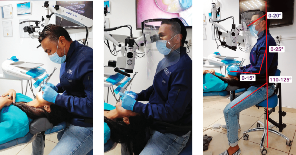
From a biomechanical point of view, the recommended neutral posture for a seated human body (Figure 2) is as follows (viewed from the side):6
- Head: Ears aligned to the shoulders, eyes on the horizon line or 0° to 20° below
- Shoulders: Aligned to the hip
- Arms: Away from the body’s midline maximum 20°; a maximum forward position from the shoulder 25°
- Forearms: Parallel to the floor, or 0° to 15° up
- Hips: Higher than knee position, 110° to 125° of angulation
- Thighs: Maximum separation of 30° aligned with the feet
The Ergonomic Stool
The ergonomic stool is a critical element for dental procedures; ergonomically designed seating will protect the pelvis, which is the foundation of seated dentistry. Considering that a dentist spends more time sitting than in bed, it is wise to invest in good ergonomic stools and pay close attention to stool characteristics. The stool should have a convex backrest that supports the concave lumbar area. Additionally, a quality seat will have a negative inclination that raises the pelvis to 110° to 125°, higher than the knees.6
Armrests can be an essential feature of an ergonomic seat. The arm’s weight is 5% of the total body weight, and support of the arms helps reduce fatigue in the upper body area, such as the shoulders and neck. Ergonomic stools come in varying designs and shapes, and the practitioner must be sure that the stool conforms to an individual’s dimensions and comfort levels before the final purchase (Figure 3).

The Dental Assistant
Four-handed dentistry is critical for good workflow and efficiency. A well-trained dental assistant will positively impact the dentist’s physical health, allowing the him or her to carry out necessary movements and focus solely and exclusively on the procedure. The assistant must control the horizontal reach distance and maintain frequently used instruments between 14 to 25 inches from the dentist at all times. This distance will allow the assistant to execute short, efficient movements and place instruments in the dentist’s hands (Figure 4).

Figure 4
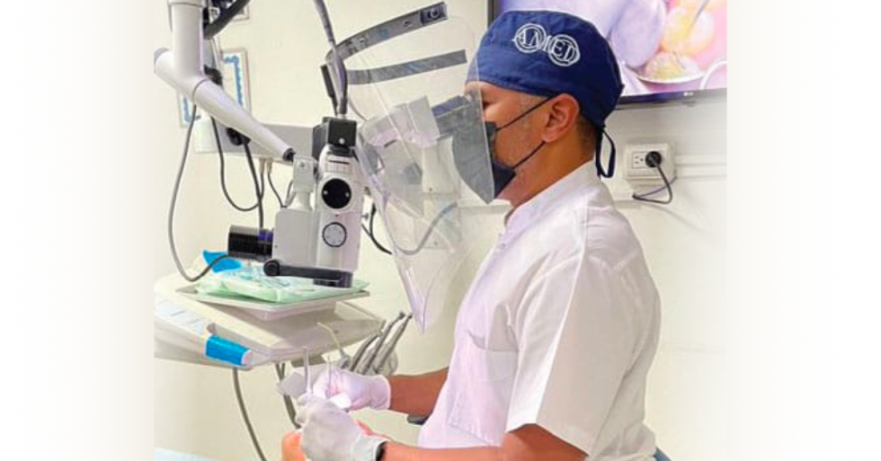
The Dental Microscope
The dental microscope can be a valuable tool for ergonomic visualization. From the biomechanical point of view, without visualization aids, such as the microscope or loupes, the operator must lean forward to see the oral cavity.7,8 The dental microscope can eliminate many elements of this problem, as it enables the dentist to gain visual information through coaxial lighting and multiple magnification steps.
With the dental microscope, the working distances allow the operator to work in a seated neutral work posture aligning the head and spine. The microscope also encourages the sitting work position at 12 o’clock, just behind the patient’s head, where postural symmetry is ideal and less pressure is put on the musculoskeletal structures.9,10 Adequate training using the dental microscope is vital to obtaining the best ergonomic benefit from the instrument.
Video and photographic benefits are also offered by the microscope. These features can allow the patient to better understand the diagnosis and procedures by visualizing the areas of concern. The video features can also aid in training the assistant.
Ultimately, these pillars are more efficient if a systematized positioning routine is implemented. The operator should begin in a seated neutral posture at 12 o’clock (in the midline of the patient’s head), followed by correctly positioning the patient in the most horizontal position possible. The oral cavity should be placed in the most strategic position for the best visual angle.
Key Takeaways
- Repetitive movement injuries can hinder a dentist’s performance and potentially end his or her career.
- The four fundamental pillars of dental ergonomics are: knowledge of the body’s biomechanics, ergonomic stool, four-handed dentistry with a dental assistant, and the use of a dental microscope.
- An ergonomic stool with proper backrest support, negative inclination, and armrests can protect the pelvis and reduce fatigue in the upper body.
- Four-handed dentistry, with a well-trained dental assistant controlling instrument reach distance, enhances workflow and efficiency.
- The dental microscope can aid in ergonomic visualization by allowing for seated neutral posture, minimizing musculoskeletal compression, and providing magnification and lighting benefits.
- Incorporating a systematic positioning routine, considering the operator’s posture, patient positioning, and use of the microscope enhances efficiency and precision in dentistry.
Conclusion
The operator’s posture is key to practicing efficiently and maintaining career longevity. Quality performance guarantees the desired therapeutic results, and relying on the correct technology and well-trained human resources makes this possible.
References
- Rising DW, Bennett BC, Hursh K, et al. Reports of body pain in a dental student population. J Am Dent Assoc. 2005;136:81–86.
- Eastman Kodak Co Ergonomic Group. Ergonomic Design for People at Work. Volume 2. New York: Van Nostrand Reinhold; 1986:21–24.
- Carter J. The inevitability of neck and back pain. Available at: dentistryiq.com/dental-hygiene/career-development/article/16348337/the-inevitability-of-neck-and-back-pain. Accessed July 18, 2023.
- Carr GB, Murgel CA. The use of the operating microscope in endodontics. Dent Clin N Am. 2010;54:191-214.
- Pîrvu C, Pătraşcu I, Pîrvu D, Ionescu C. The dentist operating posture-ergonomic aspects. J Med Life. 2014;7:177–182.
- Valachi B. Practice Dentistry Pain-Free: Evidence-Based Strategies to Prevent Pain and Extend Your Career. Portland, Oregon: Posturedontics Press; 2008;25–27.
- Bud M, Jitaru S, Lucaciu O, et al. The advantages of the dental operative microscope in restorative dentistry. Med Pharm Rep. 2021;94:22–27.
- Ortiz Hugues JC, Carlos G. Rapid upper limb assesment (RULA) and Rodgers muscle fatigue analysis (RMFA) of dentists using optical microscope, loupes or no magnification during endodontic access. A pilot study. Available at: isoes.info/conferences/2020/Papers/Hugues.pdf. Accessed July 18, 2023.
- Microscopic Dentistry: A Practical Guide. Available at: nuview.co.uk/files/ww/Microscopic%20Dentistry%20A%20Practical%20Guide.pdf. Accessed July 18, 2023.
- Blanc D, Farre P, Hamel O. Variability of musculoskeletal strain on dentists: An electromyographyc and goniometric study. Int J Occup Saf Ergon. 2014;20:295-307.
.png)