This article summarizes content published on: Updates in Clinical Dentistry written by Dr. Donato (Dino) Napoletano, a restorative dentist and long-time Global microscope user. In addition to owning and operating Donato Dental in Middletown, New York, Dr. Napoletano has conducted hands-on microscope training sessions both privately and at Tufts University’s GPR program.
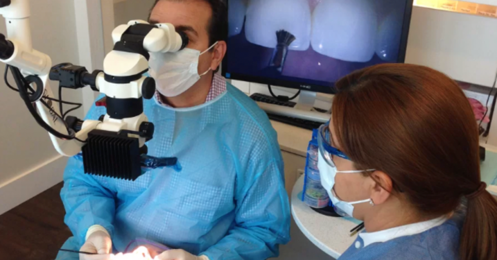
One dental technology gaining more popularity among dentists and specialists alike is a dental microscope. Although many doctors have historically viewed it as a necessity only for endodontic procedures, the DOM is beginning to gain wider acceptance.
You may find more universities incorporating dental microscopes into programs outside endodontics. It is becoming more common for undergraduate programs to feature access to this technology.
The dental microscope provides numerous benefits to doctors, including the flexibility to adjust focal distance without contorting your body, head, back or neck. This allows you to remain comfortable in a more ergonomic position.
Plus, a dental microscope dramatically increases your visibility, with multiple levels of magnification at the turn of a dial. The enhanced visibility of a dental microscope allows shadow-free lighting, thanks to a coaxial design. Unlike loupes, even with a headlamp, will create shadows in your vision.
Today, we’re going to balance the equation, to get the best visualization into the oral cavity, you need magnification + illumination. Let’s dive in.
Increased Visualization = Magnification + Illumination
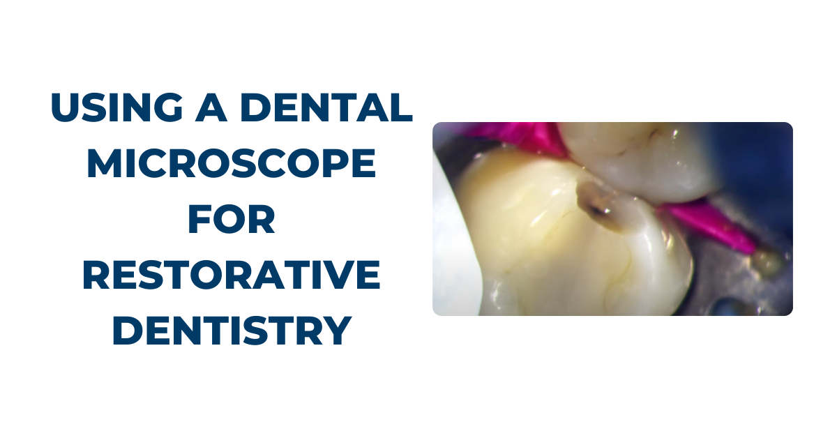
Most dental microscopes provide up to six levels of magnification, ranging from 2x to 20x (ie. 2x the magnification power of the naked eye). And the other critical component in increasing visualization is illumination.
Dental microscopes are equipped with an integrated coaxial light source that allows unobstructed, shadow-free illumination of the operating field. With coaxial illumination, the path of light is directed parallel to the optical axis, which allows significantly improved visualization of even the most difficult-to-access areas of the oral cavity.
Why is visualization important? With enhanced visualization, your ability to diagnose pathology in the earlier stages of a disease process is possible. You may find your treatments can also be performed with greater precision, thereby reducing the occurrence of failures and the need to redo procedures.
Enhanced visualization can also allow treatment to be provided more comfortably to the patient because of lighter, more refined hand movements. After training, you’ll find this may naturally start occurring when you’ve become accustomed to operating in a well-illuminated magnified field.
Ultimately, increased visualization can result in greater efficiency and productivity. You may find less time is wasted with tactile exploration and checking that all decay has been removed or that a crown margin is fully seated. When you can see all areas of the mouth or a tooth perfectly, the level of efficiency and precision in diagnosis and treatment naturally increases.
Simply put, you can’t effectively diagnose or treat what you can’t see.
Dental Operating Microscopes Versus Loupes
So, let’s put this to the test. Loupes are also popular magnification tools for many doctors looking to enhance their visualization. Doctors using loupes may be missing out on some of the enhanced visualization, as well as ergonomic benefits inherent to a dental microscope.
For full disclosure, our company manufactures microscopes, but we understand there may be areas where loupes provide advantages, such as cost. We do our best to provide honest and transparent advice so you can make an educated decision.
There are many obvious differences of dental microscopes from loupes, such as size, cost, portability, multiple levels of magnification, image stability, and the ability to integrate cameras. Less obvious but significant differences include the following:
- Reduced eye strain: Microscope users have very little eye strain and fatigue compared with loupe users, especially as magnification levels increase. This can be attributed to the difference in optical lens design between the two. Your scope relies on infinity-corrected optics that allow stereoscopic vision, whereas loupes rely on convergent optics.
- Unobstructed, shadow-free coaxial lighting: Emitted directly from the objective lens parallel to the line of vision. Lighting that is attached to loupes or a headlamp is not coaxial and, therefore, not completely shadow-free.
- Improved ergonomics: Operate in an ideal, fully upright postural position minimal to no stress or fatigue on the musculoskeletal system, especially the neck and back. Although loupes do help in improving postural ergonomics when compared with unassisted vision, some forward inclination of the head and neck is unavoidable, and the sheer weight of the loupes along with accessory lighting adds additional stressors to the musculoskeletal system.
Ready to Get Started? We’re Here to Help!
We’re Global Surgical. For over 25 years, we’ve helped doctors achieve improved visualization to benefit their practice.
We’re proud to be based in the US, with manufacturing and assembly facilities in St. Louis, MO. This helps us give our customers the best service, domestically and internationally. And, as part of our commitment to our customers, we offer a limited lifetime warranty on our scopes (US & Canada customers only).
If you’re considering adding a microscope to your practice or you just want to learn more about utilizing a dental microscope, we are here to help! Please feel free to reach out at 800-861-3585 or by clicking the button below.

.png)