Enhancing Diagnostic Capabilities & Precision: Integrating Dental Microscopes with CBCT

It’s no secret that doctors today have an overwhelming number of options when it comes to the dental equipment they’ll choose for their practice. Doctors looking to provide the highest level of care may seek to make significant investments in the dental equipment they choose. Ultimately, how you use your equipment will yield your greatest impact on how big of a return you’ll see and how quickly you’ll achieve ROI.
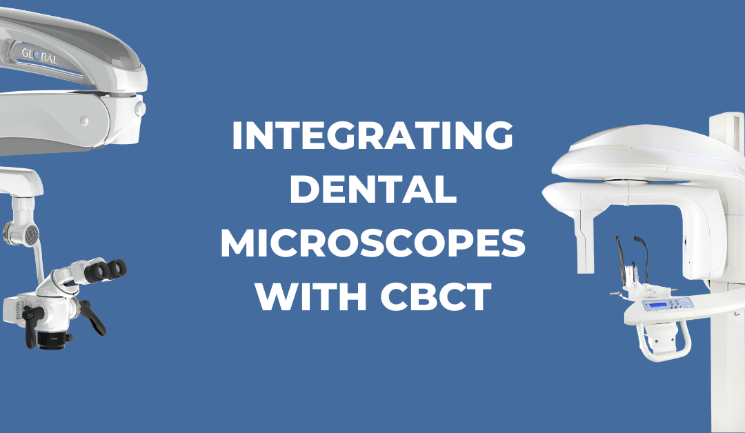
Today, we’re going to explore an emerging trend in dentistry: integration of dental microscopes with dental cone beam (CBCT). Independently, these two technologies are already commonly accepted as means to providing advanced treatment planning and enhancing diagnostic capabilities.
But, if you’re like many doctors who have made the investment in both a dental microscope and a CBCT system, have you considered how you could integrate the two?
You may already know, dental CBCT provides exceptional visualization of underlying bone structure in dental and craniofacial anatomy in 3 dimensions. And, a dental microscope enhances your vision of the oral cavity in real-time with magnification and illumination.
So, how can these two be used together? Here are 7 ways you can integrate a dental microscope with 3D imaging from a CBCT system:
- Pre-Operative Assessment: Before starting a procedure, such as endodontic treatment or implant placement, utilize dental CBCT to obtain three-dimensional images of the patient’s oral structures. CBCT provides detailed information about bone anatomy, tooth position, and neighboring structures. This information can help you identify any potential complexities or challenges in advance.
- CBCT-Guided Navigation: Use the CBCT scan data to generate surgical guides or navigation templates. These guides can be used to precisely position the dental microscope during the procedure, ensuring accurate visualization and alignment with the target area.
- Combine CBCT and Microscopic Visualization: During surgical procedures, such as root canal treatments or complex restorations, use both the dental microscope and CBCT images simultaneously. The dental microscope allows for high-magnification visualization of the treatment area, while CBCT provides additional anatomical information that may not be directly visible through the microscope. Integrating the two technologies allows for a comprehensive understanding of the patient’s dental anatomy and facilitates precise treatment execution.
- Image Fusion: Some advanced dental software systems allow for image fusion, which combines the CBCT images with the real-time microscopic view. This integration overlays the CBCT data onto the live microscope image, providing a merged view of the patient’s anatomy and the current treatment area. Image fusion can enhance the accuracy of procedures, such as implant placement, by allowing you to visualize the exact position and trajectory of the implant in relation to the patient’s bone structure.
- Real-Time Verification: Utilize the dental microscope to verify the accuracy of the treatment in real-time. As you perform the procedure, use the microscope to visually confirm the placement of instruments, fillings, or restorations based on the CBCT scan data. This verification ensures that the treatment aligns with the planned trajectory and desired outcomes.
- Documentation & Case Review: Combining the dental microscope with CBCT scans allows for comprehensive documentation and case review. Capture images or videos through the microscope during the procedure, ensuring that the recorded visuals correspond to the precise anatomical structures revealed in the CBCT scan. This documentation serves as valuable evidence for treatment planning, communication with patients, and professional collaborations.
- Enhanced Treatment Planning: The integration of a dental microscope with CBCT images improves treatment planning accuracy. By analyzing the CBCT scans in conjunction with the microscopic visualization, you can better assess the extent of pathology, evaluate canal morphology, identify hidden structures, and determine the most suitable treatment approach.
More Value With a Global Microscope
Looking to get started with a dental microscope and discover new ways to put your technology to work for you? At Global Surgical, we’re committed to your success, with durable products and our knowledgeable Technical & Customer Service teams. We’re proud to be based in the US, with manufacturing and assembly facilities in St. Louis, MO. This helps us give our customers the best service, domestically and internationally. And, as part of our commitment to our customers, we offer a limited lifetime warranty on our scopes (US & Canada customers only).
Many of the doctors we speak with begin using their scopes with nearly every patient – from observation and diagnosis to treatment planning and procedure, so we know it’s important they are able to use their scope effectively.
Questions? Reach Out!
If you’re just getting started with a dental microscope, or considering adding a scope to your practice, we are here to help! Let us help configure and customize your scope to your clinical needs, helping you get started as quickly as possible.
Get started by reaching out to us at 800-861-3585 or by clicking the button below.


