Coaxial Lighting: Illuminating Clarity in Dental Microscopy

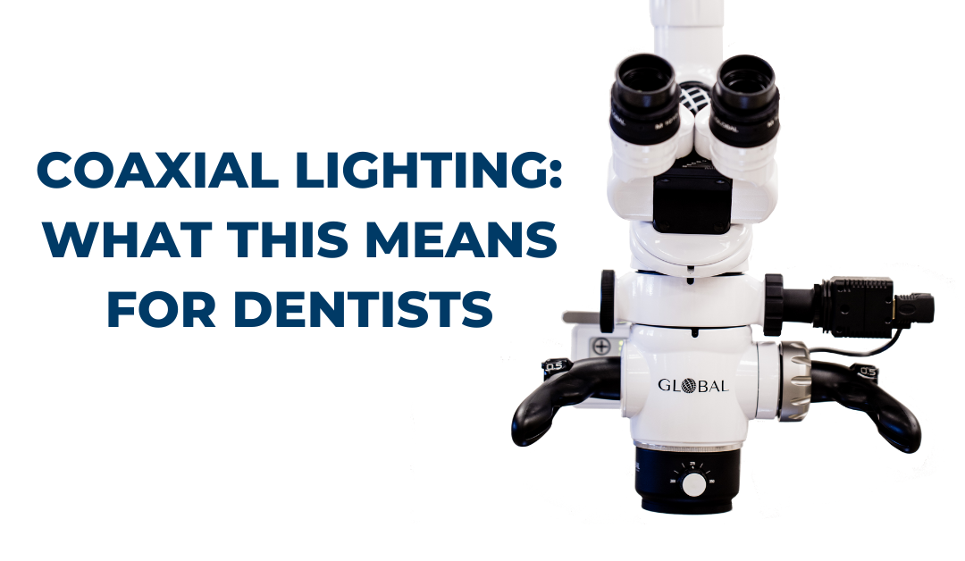
Coaxial lighting is a specific type of illumination technique used in dental microscopes to enhance visibility and improve the quality of image formation. In a dental microscope, coaxial lighting refers to the configuration where the light source is coaxially aligned with the optical axis of the microscope.
Here’s how coaxial lighting works on a dental microscope:
- Light Source: The dental microscope is equipped with a light source, typically an LED or halogen bulb, located within the microscope body or attached to it. This light source generates a beam of light that travels along the same path as the microscope’s optical axis.
- Optical Path: The light beam emitted from the light source passes through a series of optical components within the microscope, including lenses and mirrors, which help control the direction, intensity, and focus of the light.
- Beam Splitter: A beam splitter, usually a partially reflective mirror or prism, is positioned within the microscope’s optical path. It allows a portion of the light beam to pass through while reflecting the remaining portion towards the objective lens.
- Objective Lens: The objective lens, located near the tip of the microscope, is responsible for collecting and magnifying the light that interacts with the dental structures. It focuses the coaxial light onto the tooth or area of interest.
- Sample Interaction: The coaxial light illuminates the dental structure or area being examined. It interacts with the tooth surfaces, reflecting or scattering off different surfaces and structures within the oral cavity.
- Light Collection: The light that interacts with the dental structures or surrounding tissues is collected by the objective lens and directed back through the microscope’s optical system.
- Observation and Image Formation: The collected light passes through the beam splitter and is directed towards the eyepieces or camera attachment of the microscope. Dentists or operators can observe the illuminated dental structures through the eyepieces or view them on a monitor if a camera is connected.
The coaxial lighting configuration ensures that the incident light beam and the collected light beam travel along the same optical axis. This alignment minimizes shadowing and enhances visualization, as the light is directed straight onto the tooth surface being examined. It allows for detailed observation of fine details, such as cracks, fractures, or anomalies in the tooth structure.
Coaxial lighting is particularly useful in endodontic procedures, where a clear view of the root canal system is essential. The direct illumination provided by coaxial lighting helps reveal intricate canal anatomy, aiding in precise treatment planning and execution.
In summary, coaxial lighting on a dental microscope involves aligning the light source with the microscope’s optical axis to provide direct illumination onto the dental structures. This configuration minimizes shadows, enhances visualization, and allows for detailed observation of dental anatomy, contributing to improved diagnostic accuracy and treatment outcomes.
Questions? We’re Ready to Help!
If you’re considering adding a microscope to your practice or you just want to learn more about utilizing a dental microscope, we are here to help! We’ve been helping dental practices for over 25 years see the benefits of high magnification through a dental microscope.
We’re proud to be based in the US, with manufacturing and assembly facilities in St. Louis, MO. This helps us give our customers the best service, domestically and internationally. And, as part of our commitment to our customers, we offer a limited lifetime warranty on our scopes (US & Canada customers only).
Please feel free to reach out at 800-861-3585 or by clicking the button below.


.png)